Muscles that lift the Arches of the Feet
The navicular bone is found on the inner side of the foot. The navicular articulates with five of the other tarsal bones - at the top with the talus, talonavicular joint, laterally (outer side) with the cuboid, cubonavicular joint, and at the bottom it articulates with the three cuneiform bones. In around 10% of the population, a small extra piece of bone develops next to the navicular which.

Sulcus
Regions of the Foot. The foot is traditionally divided into three regions: the hindfoot, the midfoot, and the forefoot (Figure 2).Additionally, the lower leg often refers to the area between the knee and the ankle and this area is critical to the functioning of the foot.. The Hindfoot begins at the ankle joint and stops at the transverse tarsal joint (a combination of the talonavicular and.
Foot and ankle anatomy, conditions and treatments
Foot Pain Identifier. footEducation.com was created by orthopaedic surgeons to provide patients and medical. providers with current and accurate information on foot and ankle conditions and their. treatments. The contributors to this site are all board certified orthopaedic surgeons who. specialize in treating patients with foot and ankle problems.

Diagrams of Foot 101 Diagrams
Bottom Of Foot Pain Diagram. This foot pain diagram looks at the common causes of pain under the foot and at the back of the heel. A. Heel Spurs. A bone spur here is called an inferior calcaneal bone spur and is usually linked with a tight plantar fascia. Causes a sharp pain under the foot that is worse with initial movement then eases to a.

Diagram Of Your Foot
Foot. The foot is the lowermost point of the human leg. The foot's shape, along with the body's natural balance-keeping systems, make humans capable of not only walking, but also running.

Diagram Of A Human Foot Human Foot Diagram Anatomy Organ Anatomy
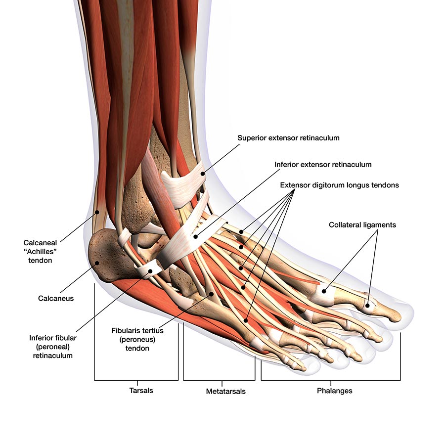
Tarsals. The tarsals are a group of seven bones close to the ankle. The proximal tarsal bones are the talus and the calcaneus, which is the largest bone of the foot. The talus is on top of the.

Diagram Of Your Foot
Listed below are 3 common areas of pain in the foot and their causes: Pain in the ball of the foot. Pain in the ball of the foot, located on the bottom of the foot behind the toes, may be caused by nerve or joint damage in that area. In addition, a benign (noncancerous) growth, such as Morton's neuroma, may cause the pain.
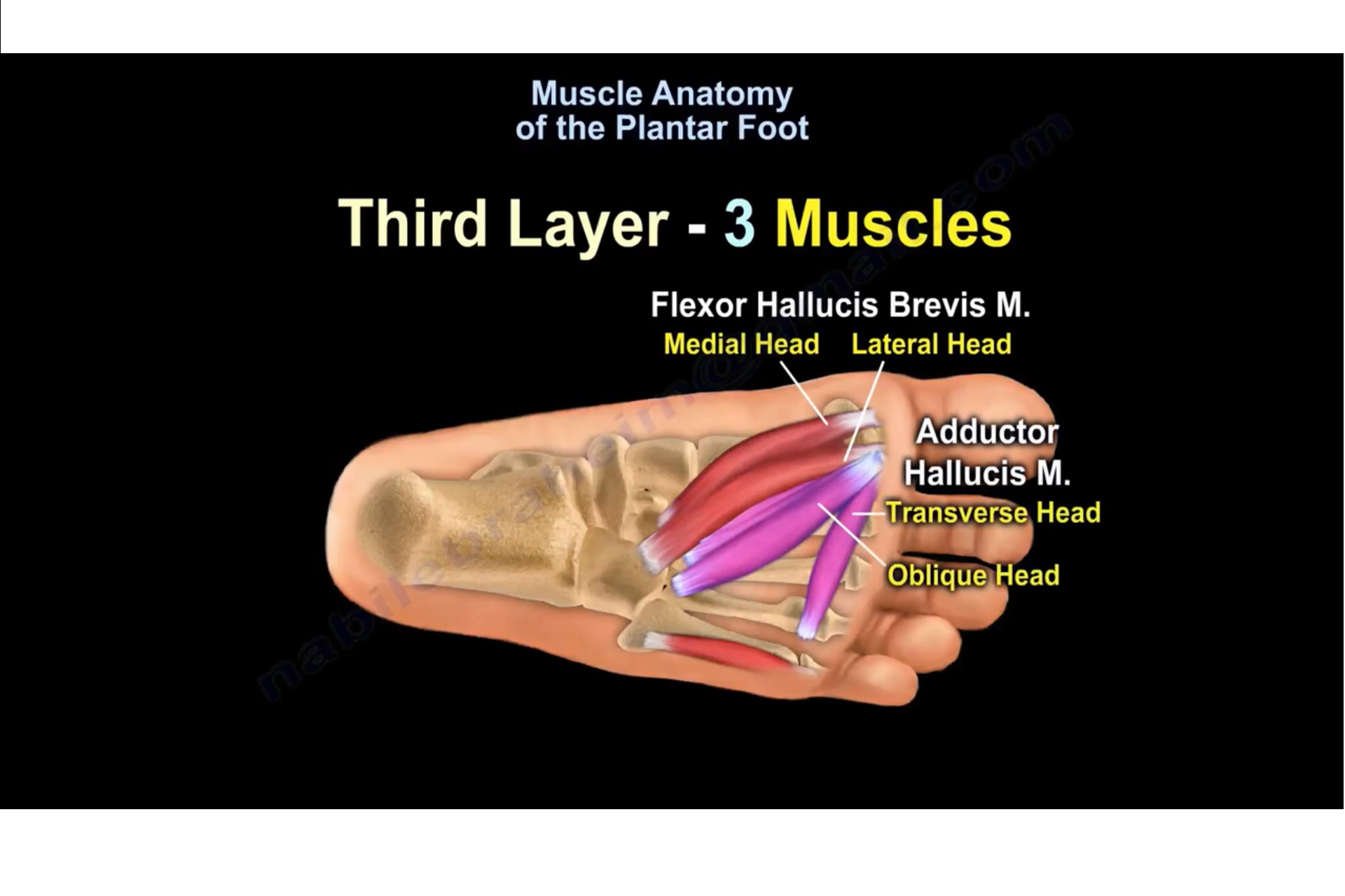
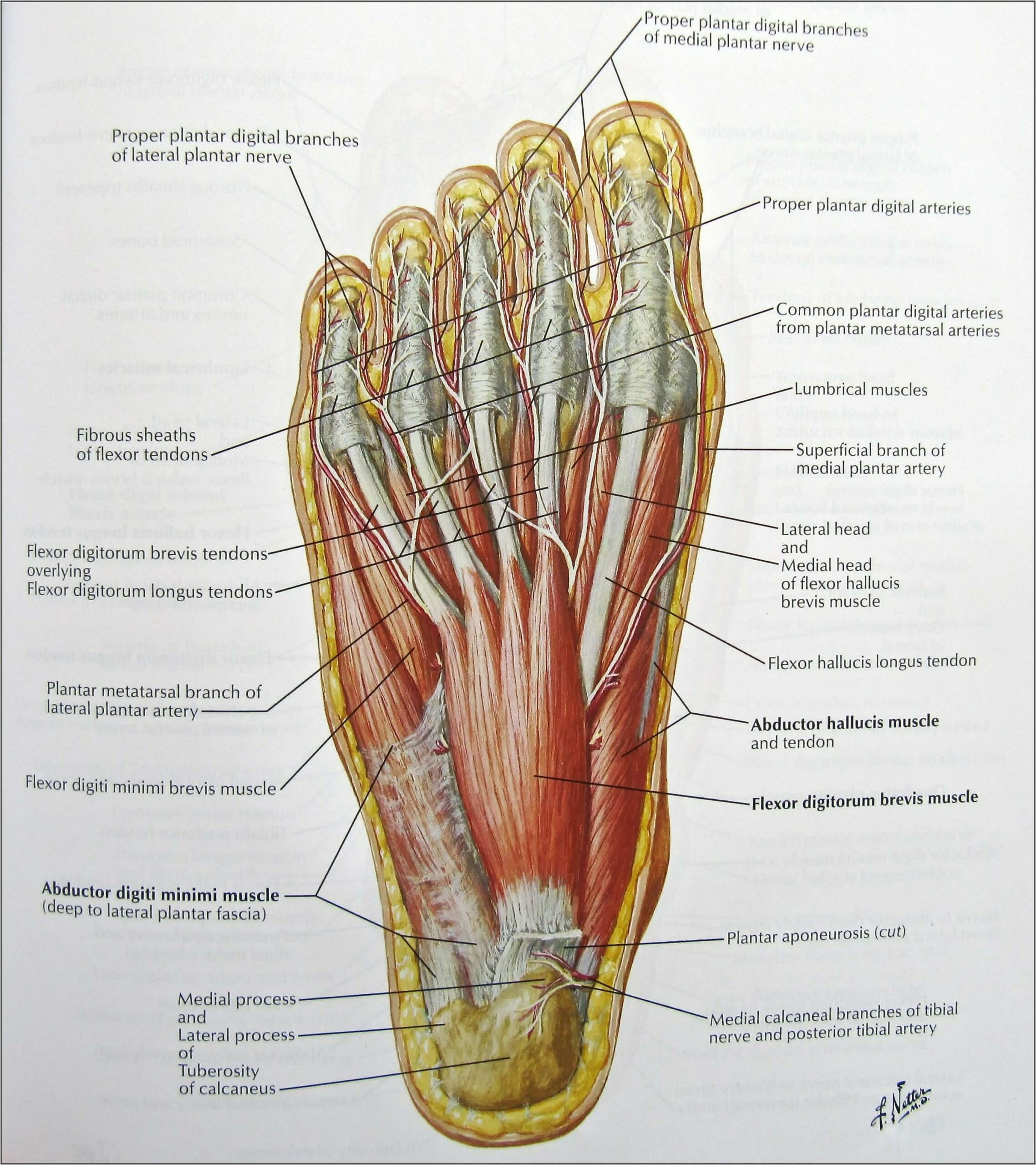
Muscle Anatomy Of The Plantar Foot —
Foot Pain Chart - Bottom of the foot. Bottom of the Heel: Fat Pad Atrophy - changes in the thickness of the fatty tissue protecting the heel bone. Spring Ligament Injury - a ligament spanning the bottom of the foot that plays a vital role alongside the Plantar Fascia to provide structural integrity to the foot.

What Is Turf Toe?
The nerves of the leg and foot arise from spinal nerves connected to the spinal cord in the lower back and pelvis. As these nerves descend toward the thighs, they form two networks of crossed nerves known as the lumbar plexus and sacral plexus. The lumbar plexus forms in the lower back from the merger of spinal nerves L1 through L4 while the.

Foot Anatomy 101 A Quick Lesson From a New Hampshire Podiatrist Nagy
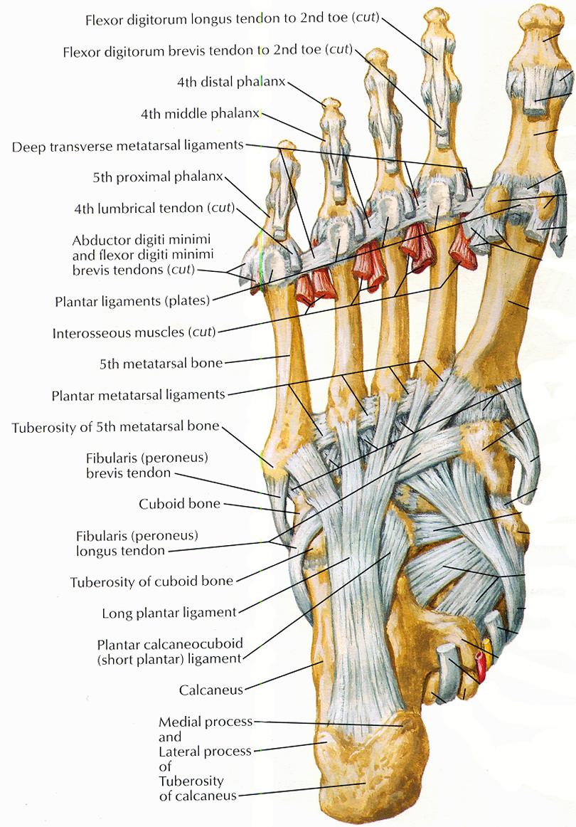
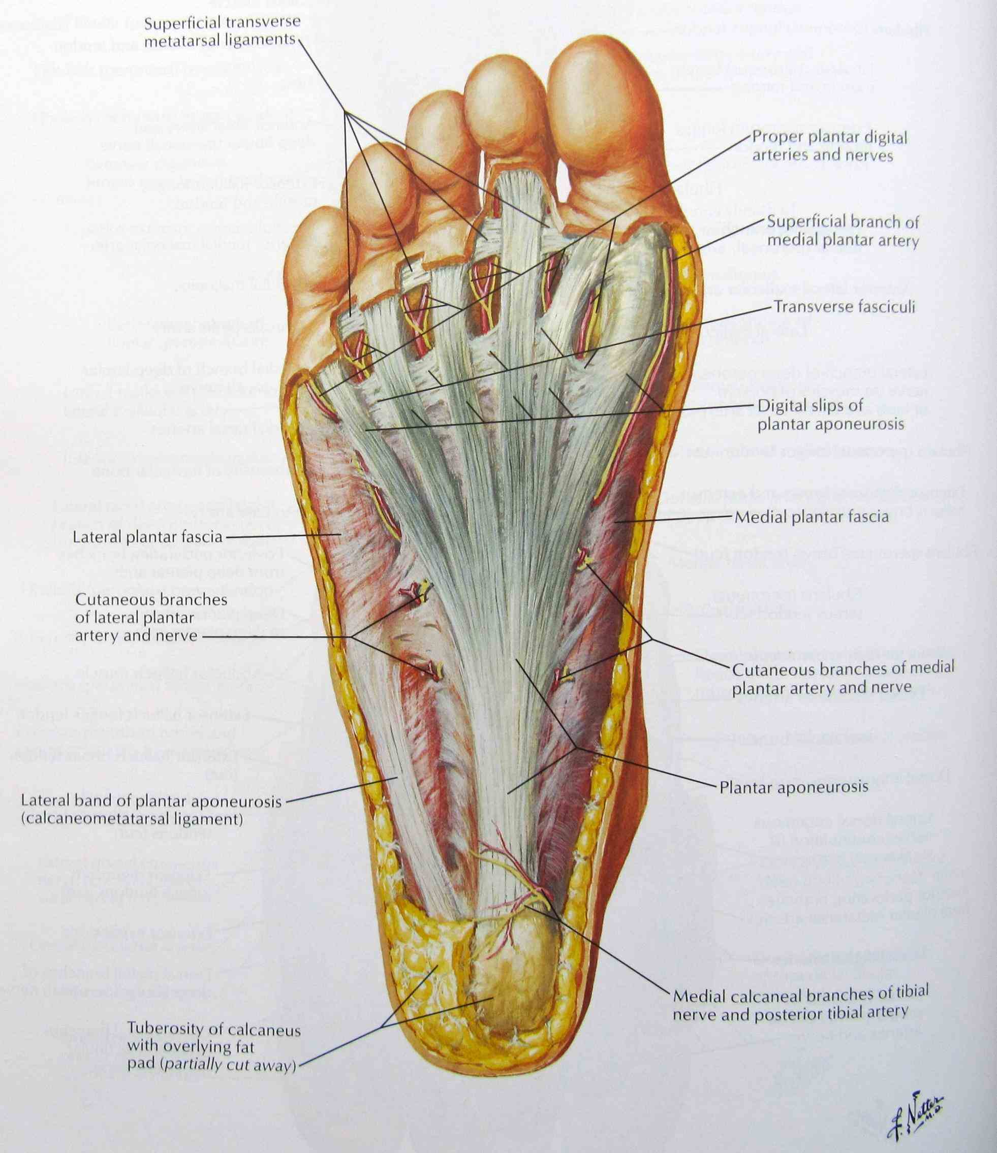
The muscles of the foot are located mainly in the sole of the foot and divided into a central (medial) group and a group on either side (lateral). The muscles at the top of the foot fan out to supply the individual toes. The tendons in the foot are thick bands that connect muscles to bones. When the muscles tighten (contract) they pull on the.

Anatomy Of The Foot Bottom Anatomy Of The Bottom Of The Foot Human
The padded area on the bottom of the foot is known as the plantar aspect. The top part of your foot above the arch is the instep. In medical terms, the top of the foot is the dorsum or dorsal region. Anatomy of the Lower Leg Muscles. Bones . There are 26 bones in the foot, and they can be categorized according to their location.

Cutaneous afferent innervation of the human foot sole what can we
Morton's Neuroma - A painful condition that most commonly affects the ball of the foot and is caused by a thickening of the nerve tissue. Commonly described as; feeling like your walking on a marble and feeling like a sock is wadded up under the foot. Metatarsal Fracture - A break or 'crack' in one of the metatarsal bones (long bones) in the.
Anatomy The Bones Of The Foot
The foot is a part of vertebrate anatomy which serves the purpose of supporting the animal's weight and allowing for locomotion on land. In humans, the foot is one of the most complex structures in the body. It is made up of over 100 moving parts - bones, muscles, tendons, and ligaments designed to allow the foot to balance the body's.

Medial Muscles And Bones Of The Foot Sole Labeled Human Anatomy Diagram
Peripheral neuropathy can cause pain on the bottom of the foot paired with tingling or burning, and so on. Finding the cause of bottom-of-the-foot pain may include a physical exam and X-rays or other imaging. Treatment may involve pain relief, lifestyle changes, physical therapy, and in severe cases, surgery. 18 Sources.
Medical Diagram Of Bottom Of Foot Diagrams Resume Template
Bones of foot. The 26 bones of the foot consist of eight distinct types, including the tarsals, metatarsals, phalanges, cuneiforms, talus, navicular, and cuboid bones. The skeletal structure of.
Bottom Of Foot Diagram Visual Diagram
The Toes, Arch and Heel. Toes are the parts of the foot that allow people to move. They help people grip the ground and push off when they walk or run. The arch is the part of the foot that helps to absorb shock when we move around. It is located between the heel and the toes. The heel provides balance and stability.
